Respiratory System Assessment Overview

Key steps include:Respiratory assessment guides nurses through systematic observation, inspection, palpation, percussion, and auscultation, emphasizing ventilation mechanics and muscle effort to overcome airway resistance. It integrates clinical context and patient history for accurate diagnosis.!!
Anatomy of the Respiratory System
The respiratory system’s structural framework is organized into the upper and lower airways, thoracic cavity, and pulmonary parenchyma. The upper airway includes the nasal cavity, pharynx, larynx, and trachea, each lined with ciliated pseudostratified columnar epithelium that facilitates mucociliary clearance. The trachea bifurcates into the mainstem bronchi, which further divide into lobar and segmental bronchi, forming a branching tree that delivers air to the alveolar sacs.
The thoracic cavity is partitioned into right and left pleural spaces separated by the mediastinum. The mediastinum houses the heart, great vessels, esophagus, and the hilum where pulmonary arteries, veins, and bronchi enter the lungs. Each pleural cavity is lined by a double‑layered pleura: the visceral pleura adheres to the lung surface, while the parietal pleura lines the thoracic wall. A thin serous fluid within the pleural space reduces friction during respiration.

- Diaphragm: the primary muscle of inspiration, dome‑shaped and attached to the xiphoid process, costal margin, and lumbar vertebrae.
- Intercostal muscles: external intercostals elevate ribs, internal intercostals depress ribs, and innermost intercostals assist in forced expiration.
- Accessory muscles: sternocleidomastoid, scalene, and pectoralis major can augment ventilation during increased demand.
- Bronchial cartilage: hyaline cartilage rings provide structural support to the trachea and bronchi.
- Alveolar ducts and sacs: terminal units where gas exchange occurs; lined by type I and type II pneumocytes.
Understanding this anatomy is essential for assessment, as variations or pathologies in these structures can alter findings and influence diagnostics and.!
Physiology of Ventilation
Ventilation is the mechanical process that moves air into and out of the lungs, driven by changes in thoracic pressure generated by the diaphragm and intercostal muscles. During inspiration, diaphragmatic contraction pulls the dome downward, expanding the thoracic cavity and decreasing intrapleural pressure. This pressure gradient draws air from the atmosphere through the airway into alveolar spaces. Expiration is typically passive; relaxation of the diaphragm and elastic recoil of the lungs and chest wall increase intrapleural pressure, forcing air out. Forced expiration requires contraction of internal intercostals and abdominal muscles, further augmenting intrathoracic pressure. The efficiency of ventilation depends on airway resistance, lung compliance, and respiratory muscle strength. Airway resistance is influenced by bronchial diameter, mucus production, and airway inflammation. Lung compliance reflects the ease with which the lung can expand; decreased compliance, as seen in fibrosis, limits tidal volume. Respiratory drive is regulated centrally by the medullary respiratory center, responding to CO₂, O₂, and pH changes detected by central and peripheral chemoreceptors. Reflexes such as coughing and sneezing protect the airway by clearing obstructions. Proper assessment of ventilation requires evaluation of breathing pattern, effort, and muscle use, which can indicate underlying pathology such as lung disease. Chemoreceptors in medulla adjust breathing to keep CO₂ normal. This assessment is vital.!!!Vital.

Assessment Components
Assessment components focus on patient history, breathing pattern, and accessory muscle use. Clinicians observe rate, rhythm, depth, and symmetry, noting any distress or abnormal effort. This holistic view guides targeted diagnostics and informs treatment planning.!
Inspection
Inspection is the initial, non‑invasive phase of a respiratory assessment, during which the clinician observes the thoracic region for symmetry, shape, and movement. The patient’s breathing pattern—rate, rhythm, depth, and effort—is noted, and any signs of respiratory distress, such as use of accessory muscles, nasal flaring, or cyanosis, are recorded. Skin color, pallor, or cyanotic discoloration may indicate oxygenation status. Chest wall expansion is assessed for equal movement; asymmetry may suggest underlying pathology. The presence of scars, surgical incisions, or deformities is noted, as these can influence ventilation mechanics. Visible tremors or tremulousness in the chest wall may reflect hyperventilation or anxiety. Posture, such as forward‑leaning or tripod position, provides clues to respiratory effort. Audible signs, such as wheezes or crackles, may be heard during inspection, offering early clues to airway obstruction or alveolar fluid accumulation; A systematic visual assessment establishes the baseline for palpation, percussion, and auscultation, ensuring a thorough evaluation of respiratory function. Additional visual cues include intercostal retractions, use of the sternocleidomastoid or scalene muscles, and the degree of subcostal retractions. The clinician may also assess the patient’s level of agitation or anxiety, which can alter breathing patterns. Documentation of these findings, often with photographic evidence, supports objective monitoring over time. Environmental factors such as ambient light, noise, and temperature can influence patient comfort and respiratory effort, and should be noted during inspection to contextualize findings. By integrating all these observations, the clinician builds a comprehensive picture of the patient’s respiratory status before proceeding to tactile and acoustic examinations. Finally, the clinician evaluates the thoracic cage for structural abnormalities, such as pectus excavatum or scoliosis, which may compromise lung expansion, and observes facial expressions and vocalization, as changes in voice quality can indicate upper airway obstruction. The clinician also notes any use of pursed‑lip breathing, which can indicate airflow limitation, and records the presence of chest wall crepitus, a potential sign of subcutaneous emphysema. All observations are recorded in the assessment sheet, allowing trend analysis over successive visits. This meticulous inspection phase is essential for identifying early signs of respiratory compromise and for tailoring the subsequent assessment steps to the patient’s specific needs. In addition, the clinician may use a visual analogue scale to quantify the patient’s perceived dyspnea during inspection, providing a subjective complement to the objective findings.
Palpation
Palpation is performed after inspection and before percussion and auscultation. The clinician places the palms of both hands on the patient’s chest wall, with fingers spread, to assess symmetry of expansion, tenderness, and the presence of tactile fremitus. The patient is asked to say “ninety-nine” or “one‑two‑three” while the clinician feels for vibrations transmitted through the lung parenchyma. A normal lung will transmit a moderate, even vibration; diminished fremitus may indicate pleural effusion, pneumothorax, or consolidation, whereas increased fremitus suggests consolidation. The clinician also palpates sternum, clavicles, margins for tenderness, crepitus, movement,assess abnormal emphysema. The use of the fingertips to feel for subcutaneous emphysema, which presents as a crackling sensation, is essential. The clinician may palpate the diaphragm by placing the hand in the epigastric region to assess its excursion during breathing, which can reveal diaphragmatic dysfunction. The clinician should assess the intercostal spaces for retractions or the use of accessory muscles, noting any paradoxical movement. The clinician should also palpate the thoracic spine for scoliosis or kyphosis, as structural deformities can impair ventilation. Documentation of findings, such as expansion and fremitus, gives a baseline for comparison during follow‑up assessments. Palpation complements visual inspection, enabling detection of subtle abnormalities not always apparent. Palpation shows chest change;
Percussion
Percussion is a tactile diagnostic technique that evaluates underlying thoracic structures by tapping the chest wall with the fingertips or the knuckles of the non‑dominant hand. The clinician places the thumb of the dominant hand on the patient’s sternum and the fingers of the other hand on the thoracic wall, then gently taps the knuckles. The resulting sound is compared bilaterally to assess symmetry and to detect abnormalities such as dullness, tympany, or hyper‑resonance. Dullness over the lung field may indicate consolidation, pleural effusion, or a mass, whereas tympany suggests an air‑filled cavity or pneumothorax. Hyper‑resonance can be seen in emphysema or over an area of decreased lung density. The technique also helps identify rib fractures, subcutaneous emphysema, or a pleural thickening. Percussion is performed in a systematic manner, starting at the apex and moving down the posterior and anterior chest, covering the entire lung field. The clinician records the quality, duration, and symmetry of the percussion note, noting any deviation from the normal resonant tone. This information, combined with inspection and palpation findings, provides a comprehensive assessment of the respiratory system and guides further diagnostic testing. Percussion is a quick, non‑invasive, and cost‑effective tool that enhances clinical decision‑making in respiratory care. Percussion helps identify lung pathology.The findings guide therapeutic interventions and monitoring.All data are logged in the patient chart now.

Auscultation
Auscultation uses a stethoscope to assess lung sounds, revealing ventilation status and airway patency. The clinician places the diaphragm on anterior, lateral, and posterior chest, moving from apex to base. Normal breath sounds—vesicular, bronchial, and bronchovesicular—are identified by frequency, intensity, and duration. Adventitious sounds such as crackles, wheezes, rhonchi, and pleural rubs indicate underlying pathology; crackles often occur during inspiration, wheezes may persist throughout. The clinician records laterality, symmetry, and any deviations from normal, correlating findings with inspection and percussion. Accurate auscultation guides diagnosis, treatment planning, and monitoring of therapeutic response in respiratory care. The stethoscope diaphragm is best for high‑frequency sounds, while the bell detects low‑frequency murmurs. Proper technique requires placing the stethoscope on the patient’s chest with firm but gentle pressure, ensuring a snug seal. The clinician listens for inspiratory and expiratory phases, noting any asymmetry. Documentation includes the location, type of sound, and any abnormal findings, which are then compared with inspection and percussion results to formulate a comprehensive assessment. During auscultation, clinicians assess for pleural friction rubs, indicating pleuritis, and for stridor. These findings, combined with vital signs, refine diagnostic accuracy and guide therapeutic decisions patient notes!!
Diagnostic Tests in PDF Resources
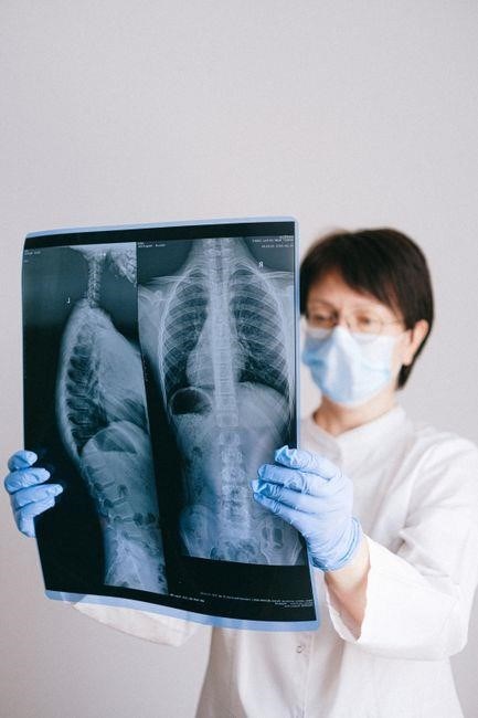
Key PDF resources detail chest X‑ray interpretation and pulmonary function tests (PFTs). The 13_CIN_Respiratory_Assessment_notes.pdf outlines image analysis, while the Respiratory System Overview PDF explains PFT parameters, reference values, and clinical significance for accurate diagnosis. ensuring accurate interpretation guiding.!!

Chest X‑Ray Interpretation
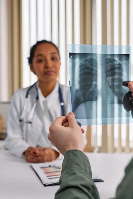
Chest X‑ray interpretation follows a systematic approach: assess image quality, position, cardiac silhouette, lung fields, trachea, diaphragmatic contour, and pleural spaces. Key findings include consolidation with air bronchograms, infiltrates, pneumothorax, effusion, atelectasis, hyperinflation, and focal masses. Correlate radiographic patterns with clinical data to guide diagnosis and management.
Interpretation of radiographic abnormalities requires recognition of patterns: consolidation suggests pneumonia or atelectasis; diffuse interstitial infiltrates may indicate pulmonary edema or fibrosis; a pleural effusion appears as blunted costophrenic angles; a pneumothorax is identified by absence of lung markings and possible mediastinal shift. Accurate assessment guides further testing and treatment.
- Consolidation: homogeneous opacity with air bronchograms, often indicating lobar pneumonia.
- Interstitial infiltrates: reticular or nodular pattern, suggestive of pulmonary edema or fibrosis.
- Pneumothorax: absence of lung markings beyond the visceral pleural line, may show mediastinal shift.
- Pleural effusion: blunted costophrenic angles, meniscus sign, size graded by radiographic extent.
These findings, when correlated with clinical presentation and laboratory data, form the basis for targeted diagnostics and therapeutic strategies, improving patient outcomes.
PDF review enhances clinical insight nowtoday now
Pulmonary Function Tests (PFTs)
Pulmonary function tests quantify airflow, lung volumes, and gas exchange, forming the cornerstone of respiratory assessment. Spirometry measures forced vital capacity (FVC) and forced expiratory volume in one second (FEV1); the FEV1/FVC ratio distinguishes obstructive (ratio <70%) from restrictive patterns (ratio normal or high with reduced volumes). Flow‑volume loops reveal characteristic flattening in obstructive disease or reduced peak flow in restrictive disease.
Lung volume assessment via body plethysmography or gas dilution determines total lung capacity (TLC) and residual volume (RV). A reduced TLC indicates restriction, whereas an elevated RV suggests air trapping. Diffusing capacity for carbon monoxide (DLCO) evaluates alveolar‑capillary transfer; decreased DLCO points to emphysema or interstitial fibrosis.
Standardization follows ATS/ERS guidelines, ensuring reproducibility. Reference equations adjust for age, sex, height, and ethnicity, allowing percent predicted values. Serial testing tracks disease progression or therapeutic response, guiding clinical decisions in asthma, COPD, and interstitial lung disease management.
In clinical practice, PFTs are interpreted against reference values that account for demographic variables, enabling clinicians to detect deviations from normal lung function. Serial testing can reveal disease progression, assess response to bronchodilators, and guide interventions, thereby improving patient outcomes soon.
Common Respiratory Pathologies Covered in PDFs
Key conditions highlighted include asthma, pneumonia, COPD, bronchiectasis, and interstitial lung disease. Each PDF outlines signs, symptoms, diagnostic criteria, and management strategies, emphasizing early detection and evidence‑based care.!!!
Asthma
Asthma is a chronic inflammatory airway disease characterized by episodic bronchoconstriction, mucus hypersecretion, and airway remodeling. Clinical presentation ranges from mild intermittent symptoms—wheeze, chest tightness, cough—to severe persistent attacks requiring intensive care. Diagnosis relies on spirometry, demonstrating reversible airflow limitation (FEV1 improvement >12% and 200 mL after bronchodilator). Peak expiratory flow variability >20% over days signals instability. Exacerbations are triggered by allergens, viral infections, cold air, exercise, or irritants, and are graded by symptom frequency and rescue inhaler use; Management follows a stepwise approach: controller inhalers (ICS, LABA combinations), rescue short‑acting β2 agonists, leukotriene modifiers, and biologics for severe eosinophilic phenotypes. Patient education on trigger avoidance, inhaler technique, and action plans is essential. Monitoring includes regular spirometry, symptom diaries, and assessment of adherence. Early intervention reduces airway hyperresponsiveness, prevents irreversible remodeling, and improves quality of life. In resource‑limited settings, peak flow meters and symptom scoring provide pragmatic assessment tools. Ongoing research explores airway microbiome, epigenetic factors, and novel biologics targeting IgE, IL‑5, and IL‑4/13 pathways, promising personalized therapy for refractory cases. Future directions emphasize precision medicine, integrating omics data and telehealth platforms to tailor interventions and improve long‑term outcomes for patientsnow!
Pneumonia
In pneumonia assessment, clinicians focus on fever, productive cough, dyspnea, and pleuritic chest pain. Physical exam reveals crackles, dullness to percussion, and decreased breath sounds. Chest X‑ray is the cornerstone, showing lobar consolidation or interstitial patterns. Laboratory tests include CBC with elevated neutrophils, CRP, and procalcitonin to gauge bacterial etiology. Sputum Gram stain and culture guide antibiotic selection. In severe cases, arterial blood gases assess hypoxemia. Point‑of‑care ultrasound detects consolidations and pleural effusions, aiding bedside diagnosis. Treatment follows pathogen‑specific guidelines: β‑lactams for typical bacteria, macrolides for atypical organisms, and fluoroquinolones for resistant strains. Supportive care includes oxygen, hydration, and antipyretics. Monitoring involves repeat imaging, vital signs, and laboratory trends to ensure resolution. Vaccination against pneumococcus and influenza reduces incidence. Early recognition and appropriate therapy prevent complications such as empyema, ARDS, and sepsis. Public health measures, including antimicrobial stewardship and infection control, are vital to curb resistance and improve outcomes. Continuous education of healthcare providers and patients about early symptom recognition and timely antibiotic use remains a cornerstone of pneumonia management. Clinicians also assess comorbidities such as chronic obstructive pulmonary disease, heart failure, and diabetes, which influence prognosis. Imaging modalities like CT scans provide detailed views of infiltrates and complications. Biomarkers including lactate dehydrogenase and interleukin‑6 help stratify severity. Follow‑up visits ensure treatment efficacy and detect relapse early. Patient education on medication adherence and symptom monitoring is vital for preventing readmissions. Regular follow‑up improves outcomes.