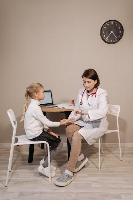
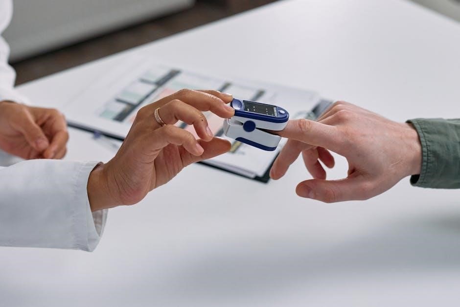
A cranial nerve examination is essential for identifying neurological conditions. It involves assessing the 12 cranial nerves, evaluating both sensory and motor functions. This process helps localize lesions, guiding further investigations and improving patient outcomes.
1.1 Importance and Purpose
Cranial nerve examination is crucial for diagnosing neurological conditions and assessing nerve function. It helps identify abnormalities, localize lesions, and guide further investigations. This systematic evaluation ensures accurate diagnosis, facilitating targeted treatment. Regular assessments aid in monitoring disease progression and recovery, making it essential for clinical practice and patient care.
1.2 Overview of the 12 Cranial Nerves
The 12 cranial nerves originate from the brain and regulate vital functions. They include olfactory (smell), optic (vision), oculomotor (eye movement), trochlear (eye rotation), trigeminal (facial sensation), abducens (eye abduction), facial (facial expression), vestibulocochlear (hearing and balance), glossopharyngeal (taste and swallowing), vagus (various visceral functions), spinal accessory (neck movement), and hypoglossal (tongue movement). Each nerve has unique roles in sensory, motor, or mixed functions, essential for maintaining bodily processes and overall health.
Preparation for Examination
Preparation involves gathering equipment, washing hands, and ensuring patient comfort. Introduce yourself, confirm identity, and explain the process. Maintain sterility and organize tools for efficient assessment.
2.1 Equipment Needed
Essential equipment includes a Snellen chart for visual acuity, a red pin for corneal reflexes, flashlight for pupillary reactions, tongue depressor for gag reflex, cotton balls for facial sensation, and olfactory stimuli. These tools enable thorough assessment of cranial nerve functions, ensuring accurate and efficient evaluation during the examination process. Ensure all tools are organized for a systematic approach.
2.2 Patient Positioning and Comfort
Position the patient comfortably, ideally sitting upright in a chair, 1 meter away for visual tests. Ensure adequate lighting for pupil reactions and eye movement assessments. Maintain a calming environment to reduce anxiety, promoting accurate responses. Proper positioning enhances the reliability of the examination, allowing for comprehensive evaluation of cranial nerve functions without patient discomfort or distraction.
Examination of Cranial Nerve I (Olfactory)
Assess the olfactory nerve by testing smell sensation using non-irritating stimuli like vanilla or coffee. Each nostril is tested separately to evaluate sensory function and detect impairments.
3.1 Testing Smell Sensation
Testing smell sensation involves using non-irritating odors like vanilla or coffee. Each nostril is occluded and tested separately. Ensure the patient identifies the odor correctly. Avoid using ammonia, as it stimulates the trigeminal nerve, providing false results. This method assesses the integrity of the olfactory pathways and detects potential impairments in smell perception effectively.
3.2 Common Abnormalities
Common abnormalities in olfactory nerve function include anosmia (loss of smell) and hyposmia (reduced smell). These may result from head trauma, infections, or tumors. Testing identifies impaired odor detection, which can indicate neurological conditions. Accurate documentation of findings is crucial for further investigation and diagnosis. Early detection aids in timely management of underlying causes, ensuring proper patient care and outcomes.

Examination of Cranial Nerve II (Optic)
The optic nerve examination assesses visual acuity, pupil reactions, and visual fields. It helps detect conditions like optic neuritis or glaucoma, ensuring early diagnosis and treatment.
4.1 Visual Acuity Testing
Visual acuity testing evaluates the sharpness of vision for each eye separately. Using Snellen charts or handheld materials, the patient reads letters or objects at a standard distance. Ensure the patient wears corrective lenses if needed. This test helps detect conditions like refractive errors or optic nerve damage. Perform this before fundoscopy to avoid dazzling the patient, ensuring accurate results.
4.2 Pupil Reactions
Pupil reactions assess the autonomic function of the optic nerve. Shine a penlight into each eye to evaluate direct and consensual responses. Note the size, symmetry, and reactivity of pupils. Abnormal findings, such as anisocoria or afferent pupillary defects, may indicate conditions like optic neuropathy or third cranial nerve lesions. This test is crucial for detecting neurological abnormalities.
Examination of Cranial Nerves III, IV, and VI (Oculomotor, Trochlear, Abducens)
Examination of cranial nerves III, IV, and VI assesses oculomotor, trochlear, and abducens functions. These nerves control eye movements and pupillary responses, crucial for diagnosing neurological conditions affecting extraocular muscles and autonomic function. Their evaluation is vital in clinical assessments.
5.1 Assessing Eye Movements
Assessing eye movements involves evaluating the function of cranial nerves III, IV, and VI. The patient is asked to follow an object with their eyes in all directions. Smooth, coordinated movements indicate normal function. Nystagmus, limited movement, or misalignment suggests nerve impairment. This test helps identify issues like strabismus or cranial nerve palsies, guiding further neurological evaluation and management.
5.2 Testing Pupil Reflexes
Testing pupil reflexes involves assessing the function of cranial nerve III. Shine a light into each eye, observing for bilateral constriction. Note any asymmetry or lack of reaction. Abnormalities, such as a Marcus Gunn pupil or anisocoria, may indicate nerve damage or central nervous system issues. This test is crucial for detecting conditions like third nerve palsy or autonomic dysfunction, aiding in early diagnosis and treatment planning.
5.3 Checking for Nystagmus
Nystagmus is an involuntary, rhythmic eye movement. During examination, ask the patient to follow a moving object with their eyes. Observe for smooth pursuit, saccadic movements, and any rhythmic oscillations. Note the direction, amplitude, and presence of a null point. Nystagmus may indicate vestibular dysfunction, brainstem lesions, or neurological disorders. Document findings carefully, as they aid in diagnosing underlying conditions affecting cranial nerves III, IV, and VI.

Examination of Cranial Nerve V (Trigeminal)
Assess the trigeminal nerve by testing facial sensation with cotton and evaluating motor function by palpating the masseter muscles. Avoid irritating substances like ammonia during testing.
6.1 Testing Facial Sensation
Testing facial sensation involves lightly touching the face with a soft object like cotton to assess sensitivity. Compare bilateral responses to detect asymmetry. Ensure not to use irritating substances that may stimulate the trigeminal nerve involuntarily. This method helps identify sensory deficits or hyperesthesia, guiding neurological assessment and diagnosis of potential nerve damage or pathology.
6.2 Assessing Motor Function
Assessing motor function involves evaluating the trigeminal nerve’s role in controlling facial muscles. Lightly brush the patient’s face or apply a soft stimulus to observe bilateral responses. Inspect for facial asymmetry or weakness. Ensure not to use irritating substances like ammonia, as they may trigger involuntary reflexes. This test helps identify motor deficits, assisting in diagnosing conditions affecting the trigeminal nerve’s motor pathways.
Examination of Cranial Nerve VII (Facial)
The facial nerve controls facial muscles and taste. Assess muscle strength by observing facial expressions and symmetry. Evaluate motor function to detect weakness or asymmetry, aiding in identifying nerve impairment.
7.1 Testing Facial Muscle Strength
Testing facial muscle strength involves assessing the patient’s ability to perform specific facial movements. Ask the patient to smile, frown, and show teeth to evaluate symmetry and strength. Observe for drooping or weakness, which may indicate facial nerve impairment. Ensure the patient’s eyes are closed tightly to check for incomplete closure, a sign of potential dysfunction. This step helps identify motor deficits accurately.
7.2 Assessing Taste Sensation
Assessing taste sensation involves using specific stimuli to evaluate the patient’s ability to detect sweetness, sourness, saltiness, and bitterness. Apply taste strips or solutions to the patient’s tongue, ensuring the areas innervated by the facial nerve are tested. Record the patient’s responses to determine if there’s any impairment in taste perception, which could indicate a potential issue with the facial nerve’s function.

Examination of Cranial Nerve VIII (Vestibulocochlear)
The vestibulocochlear nerve examination evaluates hearing and balance. Use tuning forks or audiometry for hearing assessment and balance tests like Romberg or caloric reflex to identify abnormalities.
8.1 Hearing Assessment
Hearing assessment for cranial nerve VIII involves evaluating both air and bone conduction. Techniques include pure-tone audiometry, speech recognition tests, and tympanometry. Use of tuning forks, such as Weber and Rinne tests, helps differentiate conductive from sensorineural hearing loss. Ensure a quiet environment and proper calibration of equipment for accurate results, documenting any abnormalities for further referral.
8.2 Balance Testing
Balance testing evaluates the vestibular component of cranial nerve VIII. Techniques include the Romberg test, Dix-Hallpike maneuver, and vestibular-ocular reflex assessment. These tests detect impairments in equilibrium and vestibular function. Abnormal findings may indicate vestibular nerve damage or central nervous system disorders, guiding further diagnostic steps and management strategies.

Examination of Cranial Nerves IX, X, XI (Glossopharyngeal, Vagus, Spinal Accessory)
This section focuses on assessing the glossopharyngeal, vagus, and spinal accessory nerves. It covers testing the gag reflex, evaluating speech patterns, and examining shoulder movement to identify potential deficits.
9.1 Testing Gag Reflex
Testing the gag reflex involves gently stimulating the posterior pharynx or tonsillar pillars with a tongue depressor. A normal response is elevation of the uvula and contraction of the pharyngeal muscles. Absence or asymmetry may indicate glossopharyngeal or vagus nerve dysfunction. Ensure patient is relaxed and breathing comfortably during the test to avoid false negatives or discomfort.
9.2 Assessing Speech and Swallowing
Assess speech by asking the patient to repeat phrases, observing articulation, fluency, and voice quality. Swallowing is evaluated by having the patient drink water and observing for signs of dysphagia, such as coughing or choking. These functions are primarily mediated by the vagus nerve (CN X) and glossopharyngeal nerve (CN IX). Abnormal findings may indicate neurological or structural impairments.
9.3 Evaluating Shoulder Movement
Evaluate shoulder movement by assessing the strength and symmetry of shrugging shoulders against resistance. This tests the spinal accessory nerve (CN XI), which innervates the trapezius and sternocleidomastoid muscles. Observe for winging of the scapula during arm movements. Weakness or asymmetry may indicate nerve damage or dysfunction, often seen in traumatic injuries or neurological conditions affecting CN XI.

Examination of Cranial Nerve XII (Hypoglossal)
Examine cranial nerve XII by assessing tongue protrusion, movement, and strength. Look for deviation, atrophy, or fasciculations, which may indicate nerve damage or neurological conditions.
10.1 Assessing Tongue Movement
Assessing tongue movement involves observing protrusion, lateral deviation, and strength. Instruct the patient to stick out their tongue and move it side-to-side. Note any weakness, atrophy, or tremors, which may indicate hypoglossal nerve dysfunction. Ensure the tongue protrudes midline without deviation, as asymmetry suggests nerve damage. Document any fasciculations or abnormal movements for further evaluation.
Special Tests in Cranial Nerve Examination
Special tests enhance diagnostic accuracy by targeting specific cranial nerve functions. These include visual field testing, olfactory assessments, and reflex evaluations, aiding in detecting subtle abnormalities and guiding further investigations.
11.1 Visual Field Testing
Visual field testing assesses peripheral and central vision, crucial for detecting defects caused by optic nerve damage or conditions like glaucoma. Techniques include confrontation testing, tangent screen exams, or automated perimetry. These methods help identify scotomas, hemianopsias, or other field defects, providing insights into neurological or ophthalmological conditions. Accurate results guide further investigations and management, ensuring early detection of potential issues affecting vision and neurological function.
11.2 Olfactory Testing with Specific Stimuli
Olfactory testing uses specific stimuli like essential oils or odorants to assess smell sensation. Each nostril is tested separately, ensuring the patient identifies the scent. This method helps differentiate between anosmia and hyposmia, providing insights into olfactory nerve function. Abnormal results may indicate cranial nerve I impairment or conditions like sinusitis. Accurate testing aids in diagnosing underlying pathologies affecting the sense of smell.

Documentation and Reporting
Documenting cranial nerve findings involves clear, concise notes on normal and abnormal results. Use structured PDF formats for organization, including images and diagrams to support observations, ensuring comprehensive reporting.
12.1 Structuring Findings in a PDF Format
Structuring cranial nerve examination findings in a PDF format ensures clarity and organization. Include sections for each nerve, noting normal and abnormal results. Use bullet points for easy readability and incorporate tables or charts to summarize data. Add images or diagrams to visually support findings, enhancing the report’s comprehensiveness and facilitating quick reference for healthcare providers.
12.2 Including Images and Diagrams
Including images and diagrams in a cranial nerve examination PDF enhances clarity and understanding. Use labeled illustrations of nerve pathways, anatomical structures, and abnormal findings; Incorporate visual aids like flowcharts or comparison tables to highlight normal versus pathological results. This visual support simplifies complex information, making the report more accessible for both healthcare professionals and educational purposes.

Common Pathologies and Implications
Cranial nerve pathologies include conditions like trigeminal neuralgia, facial nerve palsy, and optic neuritis. Early detection through examination is crucial for timely intervention and improved patient outcomes.
13.1 Recognizing Abnormal Findings
Abnormal findings in cranial nerve examination include loss of function, such as reduced sensation, weakness, or impaired reflexes. Signs like nystagmus, diplopia, or facial asymmetry may indicate pathology. These findings help identify specific nerve involvement and guide further diagnostic steps, ensuring appropriate management and referral for conditions like trigeminal neuralgia or optic neuritis.
13.2 Referral and Further Investigations
When abnormal findings are detected, referral to a specialist, such as a neurologist, is crucial. Further investigations like MRI, CT scans, or electrophysiological tests may be needed to confirm diagnoses. Timely referral ensures appropriate management of conditions, such as cranial nerve palsies or neuropathies, improving patient outcomes and reducing complications associated with delayed treatment.
A thorough cranial nerve examination is vital for accurate diagnoses. Key points include systematic assessment, patient history, and clear documentation. Best practices involve using mnemonics, ensuring patient comfort, and maintaining precise records for effective communication and further investigations.
14.1 Summary of Key Points
A cranial nerve examination requires a systematic approach, assessing each nerve’s sensory and motor functions. Recognizing abnormalities, such as impaired smell or vision, is crucial. Proper documentation ensures clear communication and guides further investigations. Regular practice and updated knowledge enhance examination accuracy, aiding in timely diagnoses and effective patient care.
14.2 Tips for Effective Examination
Ensure a structured approach, starting with introductions and patient comfort. Use visual aids to clarify tests. Avoid rushing to prevent missing subtle abnormalities. Maintain organization of equipment and notes. Practice regularly to improve efficiency and accuracy. Consider patient-specific factors like vision or hearing impairments. Clear communication enhances cooperation and examination outcomes.


